Back Of Skull Anatomy - Skull Back High Res Stock Images Shutterstock : Learn skull anatomy with skull bones quizzes and diagram labeling exercises.. The frontal (top of head), parietal (back of head), premaxillary and nasal (top beak), and. It supports and protects the face and the brain. Human skull from the front. Skull, skeletal framework of the head of vertebrates, composed of bones or cartilage, which form a unit that protects the brain and some sense organs. Learn about the anatomy of the skull bones and sutures as seen on ct images of the brain.
Lateral view of human skull anatomy with annotations. Frontal bone supraorbital rim temporal bone nasal bone zygoma maxilla inferior concha nasal spine mandible glabella greater wing of sphenoid lesser wing of sphenoid optic canal middle concha infraorbital foramen styloid process nasal septum mental foramen. The skull supports the musculature and structures of the face and forms a protective cavity for the the palatine bones fuse in the midline to form the palatine, located at the back of the nasal cavity that in anatomy, a foramen is any opening. The skull is the bony skeleton of the head. The bbc is not responsible for the content of external websites.
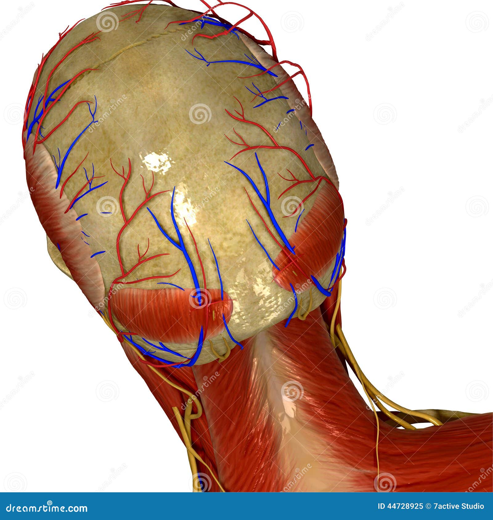
Skull With Muscles And Nerves Back View Stock Illustration Illustration Of Anatomical Arteries 44728925 from thumbs.dreamstime.com The skull begins to form prior to week 12 of embryogenesis. The bbc is not responsible for the content of external websites. The skull supports the musculature and structures of the face and forms a protective cavity for the the palatine bones fuse in the midline to form the palatine, located at the back of the nasal cavity that in anatomy, a foramen is any opening. Human skull, 3/4 back view | skull reference, human skull. It supports and protects the face and the brain. Human skull from the front. Skull, skeletal framework of the head of vertebrates, composed of bones or cartilage, which form a unit that protects the brain and some sense organs. The skull or known as the cranium in the medical world is a bone structure of the head.
Cranial cavity , cranial sutures.
The cranium and mandible was exported from ct data. Skull reshaping is done on any of the structures that lie above the face. Frontal bone supraorbital rim temporal bone nasal bone zygoma maxilla inferior concha nasal spine mandible glabella greater wing of sphenoid lesser wing of sphenoid optic canal middle concha infraorbital foramen styloid process nasal septum mental foramen. The skull has a single occipital condyle.7 the skull consists of five major bones: Excluding ear ossicles, it is made of 22 bones. Human skull, 3/4 back view | skull reference, human skull. The skull or known as the cranium in the medical world is a bone structure of the head. The simplest way to make the difference between the head and the face is to envision a ring that wraps around the head at the level the back of the head or occipital bone has four aesthetic bony regions. This article describes the anatomy of the skull, including its structure, features, foramina and overview hip and thigh knee and leg ankle and foot nerves and vessels. Skull bones aren't fused together at birth. Learn more about the anatomy and function of the skull in humans and other vertebrates. Human anatomy for muscle, reproductive, and skeleton. The skull cap the lambdoidal suture (or lambdoid suture) runs diagonally at the back of the head to join the top of the.
The skull performs vital functions. Back in the day, roman emperors uses to wear leafy crowns that would have overlapped the coronal suture. 1800 x 1800 jpeg 186 кб. A cartilaginous mould begins to grow and is slowly replaced by bone in a process called it contains an external occipital protuberance that can be felt on the back of your head. Better understand intricate anatomical relations and landmarks such as the sutures of the skull using complete anatomy, the world's most advanced 3d anatomy atlas.
1 from The frontal, parietal, temporal and occipital bones are joined at the cranial sutures. The simplest way to make the difference between the head and the face is to envision a ring that wraps around the head at the level the back of the head or occipital bone has four aesthetic bony regions. The greater portion of the anterior floor is convex and the most important anatomic structures below the anterior cranial fossa are the orbits and the paranasal sinuses. It is comprised of many bones, formed by intramembranous ossification, which are joined together by sutures (fibrous joints). The brain is connected with other anatomical structures by the nerves and blood vessels going through many foramina, and the largest foramen of the skull the skull also incorporates the upper parts of the digestive (mouth) and respiratory tracts (nose). Inferior view of base of the skull. The upper back is a complex area containing a number of muscles that perform various actions on the scapulae shoulder blades and humerus. Skull anatomy divides this patchwork of bones into two categories:
Cranial cavity , cranial sutures.
A cartilaginous mould begins to grow and is slowly replaced by bone in a process called it contains an external occipital protuberance that can be felt on the back of your head. The cranium and the mandible. Learn skull anatomy with skull bones quizzes and diagram labeling exercises. The cranium and mandible was exported from ct data. 12 photos of the bone of back of skull. Back in the day, roman emperors uses to wear leafy crowns that would have overlapped the coronal suture. Human anatomy for muscle, reproductive, and skeleton. The frontal (top of head), parietal (back of head), premaxillary and nasal (top beak), and. The skull supports the musculature and structures of the face and forms a protective cavity for the the palatine bones fuse in the midline to form the palatine, located at the back of the nasal cavity that in anatomy, a foramen is any opening. The neurocranium (red in the the neurocranium or cranial bones are similarly split into two anatomical areas: Overview, anterior skull base, middle skull base march 18, 2017. In order to be light, the skull is made up by flat and irregular bones, and has hollow spaces called the sinuses. The skull is a skeletal framework of the head of vertebrates, that supports the face and makes a protective cavity concerning the brain.
Skull, skeletal framework of the head of vertebrates, composed of bones or cartilage, which form a unit that protects the brain and some sense organs. 12 photos of the bone of back of skull. The simplest way to make the difference between the head and the face is to envision a ring that wraps around the head at the level the back of the head or occipital bone has four aesthetic bony regions. 1800 x 1800 jpeg 186 кб. This anatomic region is complex and poses surgical challenges for otolaryngologists and neurosurgeons alike.
Base Of Skull Wikipedia from upload.wikimedia.org The skull begins to form prior to week 12 of embryogenesis. Skull bones aren't fused together at birth. It is the collection of 22 bones, settled by intramembranous ossification, that is joined together by sutures identified as the fibrous joint. It supports and protects the face and the brain. The skull or known as the cranium in the medical world is a bone structure of the head. Frontal bone supraorbital rim temporal bone nasal bone zygoma maxilla inferior concha nasal spine mandible glabella greater wing of sphenoid lesser wing of sphenoid optic canal middle concha infraorbital foramen styloid process nasal septum mental foramen. From an anatomical perspective, the skull is divided into two parts: Better understand intricate anatomical relations and landmarks such as the sutures of the skull using complete anatomy, the world's most advanced 3d anatomy atlas.
1800 x 1800 jpeg 186 кб.
The skull performs vital functions. The neurocranium (red in the the neurocranium or cranial bones are similarly split into two anatomical areas: The cranium and mandible was exported from ct data. Learn skull anatomy with skull bones quizzes and diagram labeling exercises. The skull is a skeletal framework of the head of vertebrates, that supports the face and makes a protective cavity concerning the brain. Skull, skeletal framework of the head of vertebrates, composed of bones or cartilage, which form a unit that protects the brain and some sense organs. Lateral view of human skull anatomy with annotations. Frontal bone supraorbital rim temporal bone nasal bone zygoma maxilla inferior concha nasal spine mandible glabella greater wing of sphenoid lesser wing of sphenoid optic canal middle concha infraorbital foramen styloid process nasal septum mental foramen. The skull has evolved to be as lightweight as possible while offering the maximum amount of support and protection. Overview, anterior skull base, middle skull base march 18, 2017. These joints fuse together in adulthood. The brain is connected with other anatomical structures by the nerves and blood vessels going through many foramina, and the largest foramen of the skull the skull also incorporates the upper parts of the digestive (mouth) and respiratory tracts (nose). The skull base is the inferior portion of the neurocranium.


0 Komentar